✨ New Arrivals Just Dropped!Explore

Nolte's The Human Brain in Photographs and Diagrams, 5e
Pages: 294,
Specialty: Neuroanatomy,
Publisher: Elsevier,
Publication Year: 2019,
Cover: Paperback,
Dimensions: 216x276x12mm
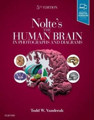
In the 5th Edition of this highly accessible atlas, Dr. Todd Vanderah continues the mission of his esteemed colleague, Dr. John "Jack" Nolte, to clearly depict and explain the challenging subject of neuroanatomy. Designed to promote a rapid understanding of complex concepts, Nolte's The Human Brain in Photographs and Diagrams combines easy-to-digest coverage of the brain, spinal cord, and brainstem with carefully selected visuals to cover all aspects of the information needed for success in coursework, on exams, and in clerkships and clinical practice. Features more than 600 high-quality figures including brain sections (transverse, coronal, axial, sagittal), 3-D reconstructions, MRIs and angiography, illustrated pathways that help you visualize anatomical structures and neuropathology. Presents a systemic series of unlabelled whole brain sections next to corresponding sections with important structures outlined and labelled. Includes a NEW chapter: An Introduction to Neuropathology, as well as NEW review questions online. Helps you understand the connections between functional systems with detailed diagrams that incorporate actual brain and spinal cord sections. Features clinical content throughout that shows how neuroanatomy applies to clinical practice. Discusses every labelled structure in the highly illustrated glossary at the end of the book. Shows major structures and major transitions in higher magnification for greater detail, and features bold index entries to indicate particularly clear illustrations of a given structure. Enhanced eBook version included with purchase. Your enhanced eBook allows you to access all of the text, figures, and references - including 68 bonus dissection videos - from the book on a variety of devices. show more
Specialty: Neuroanatomy,
Publisher: Elsevier,
Publication Year: 2019,
Cover: Paperback,
Dimensions: 216x276x12mm
In the 5th Edition of this highly accessible atlas, Dr. Todd Vanderah continues the mission of his esteemed colleague, Dr. John "Jack" Nolte, to clearly depict and explain the challenging subject of neuroanatomy. Designed to promote a rapid understanding of complex concepts, Nolte's The Human Brain in Photographs and Diagrams combines easy-to-digest coverage of the brain, spinal cord, and brainstem with carefully selected visuals to cover all aspects of the information needed for success in coursework, on exams, and in clerkships and clinical practice. Features more than 600 high-quality figures including brain sections (transverse, coronal, axial, sagittal), 3-D reconstructions, MRIs and angiography, illustrated pathways that help you visualize anatomical structures and neuropathology. Presents a systemic series of unlabelled whole brain sections next to corresponding sections with important structures outlined and labelled. Includes a NEW chapter: An Introduction to Neuropathology, as well as NEW review questions online. Helps you understand the connections between functional systems with detailed diagrams that incorporate actual brain and spinal cord sections. Features clinical content throughout that shows how neuroanatomy applies to clinical practice. Discusses every labelled structure in the highly illustrated glossary at the end of the book. Shows major structures and major transitions in higher magnification for greater detail, and features bold index entries to indicate particularly clear illustrations of a given structure. Enhanced eBook version included with purchase. Your enhanced eBook allows you to access all of the text, figures, and references - including 68 bonus dissection videos - from the book on a variety of devices. show more
Product Information
Product Information
Shipping & Returns
Shipping & Returns






Nolte's The Human Brain in Photographs and Diagrams, 5e
Nolte's The Human Brain in Photographs and Diagrams, 5e
Pages: 294,
Specialty: Neuroanatomy,
Publisher: Elsevier,
Publication Year: 2019,
Cover: Paperback,
Dimensions: 216x276x12mm
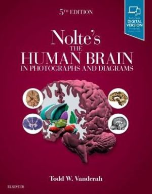
In the 5th Edition of this highly accessible atlas, Dr. Todd Vanderah continues the mission of his esteemed colleague, Dr. John "Jack" Nolte, to clearly depict and explain the challenging subject of neuroanatomy. Designed to promote a rapid understanding of complex concepts, Nolte's The Human Brain in Photographs and Diagrams combines easy-to-digest coverage of the brain, spinal cord, and brainstem with carefully selected visuals to cover all aspects of the information needed for success in coursework, on exams, and in clerkships and clinical practice. Features more than 600 high-quality figures including brain sections (transverse, coronal, axial, sagittal), 3-D reconstructions, MRIs and angiography, illustrated pathways that help you visualize anatomical structures and neuropathology. Presents a systemic series of unlabelled whole brain sections next to corresponding sections with important structures outlined and labelled. Includes a NEW chapter: An Introduction to Neuropathology, as well as NEW review questions online. Helps you understand the connections between functional systems with detailed diagrams that incorporate actual brain and spinal cord sections. Features clinical content throughout that shows how neuroanatomy applies to clinical practice. Discusses every labelled structure in the highly illustrated glossary at the end of the book. Shows major structures and major transitions in higher magnification for greater detail, and features bold index entries to indicate particularly clear illustrations of a given structure. Enhanced eBook version included with purchase. Your enhanced eBook allows you to access all of the text, figures, and references - including 68 bonus dissection videos - from the book on a variety of devices. show more
Specialty: Neuroanatomy,
Publisher: Elsevier,
Publication Year: 2019,
Cover: Paperback,
Dimensions: 216x276x12mm
In the 5th Edition of this highly accessible atlas, Dr. Todd Vanderah continues the mission of his esteemed colleague, Dr. John "Jack" Nolte, to clearly depict and explain the challenging subject of neuroanatomy. Designed to promote a rapid understanding of complex concepts, Nolte's The Human Brain in Photographs and Diagrams combines easy-to-digest coverage of the brain, spinal cord, and brainstem with carefully selected visuals to cover all aspects of the information needed for success in coursework, on exams, and in clerkships and clinical practice. Features more than 600 high-quality figures including brain sections (transverse, coronal, axial, sagittal), 3-D reconstructions, MRIs and angiography, illustrated pathways that help you visualize anatomical structures and neuropathology. Presents a systemic series of unlabelled whole brain sections next to corresponding sections with important structures outlined and labelled. Includes a NEW chapter: An Introduction to Neuropathology, as well as NEW review questions online. Helps you understand the connections between functional systems with detailed diagrams that incorporate actual brain and spinal cord sections. Features clinical content throughout that shows how neuroanatomy applies to clinical practice. Discusses every labelled structure in the highly illustrated glossary at the end of the book. Shows major structures and major transitions in higher magnification for greater detail, and features bold index entries to indicate particularly clear illustrations of a given structure. Enhanced eBook version included with purchase. Your enhanced eBook allows you to access all of the text, figures, and references - including 68 bonus dissection videos - from the book on a variety of devices. show more
$19.42
Original: $55.50
-65%Nolte's The Human Brain in Photographs and Diagrams, 5e—
$55.50
$19.42Product Information
Product Information
Shipping & Returns
Shipping & Returns
Description
Pages: 294,
Specialty: Neuroanatomy,
Publisher: Elsevier,
Publication Year: 2019,
Cover: Paperback,
Dimensions: 216x276x12mm
In the 5th Edition of this highly accessible atlas, Dr. Todd Vanderah continues the mission of his esteemed colleague, Dr. John "Jack" Nolte, to clearly depict and explain the challenging subject of neuroanatomy. Designed to promote a rapid understanding of complex concepts, Nolte's The Human Brain in Photographs and Diagrams combines easy-to-digest coverage of the brain, spinal cord, and brainstem with carefully selected visuals to cover all aspects of the information needed for success in coursework, on exams, and in clerkships and clinical practice. Features more than 600 high-quality figures including brain sections (transverse, coronal, axial, sagittal), 3-D reconstructions, MRIs and angiography, illustrated pathways that help you visualize anatomical structures and neuropathology. Presents a systemic series of unlabelled whole brain sections next to corresponding sections with important structures outlined and labelled. Includes a NEW chapter: An Introduction to Neuropathology, as well as NEW review questions online. Helps you understand the connections between functional systems with detailed diagrams that incorporate actual brain and spinal cord sections. Features clinical content throughout that shows how neuroanatomy applies to clinical practice. Discusses every labelled structure in the highly illustrated glossary at the end of the book. Shows major structures and major transitions in higher magnification for greater detail, and features bold index entries to indicate particularly clear illustrations of a given structure. Enhanced eBook version included with purchase. Your enhanced eBook allows you to access all of the text, figures, and references - including 68 bonus dissection videos - from the book on a variety of devices. show more
Specialty: Neuroanatomy,
Publisher: Elsevier,
Publication Year: 2019,
Cover: Paperback,
Dimensions: 216x276x12mm
In the 5th Edition of this highly accessible atlas, Dr. Todd Vanderah continues the mission of his esteemed colleague, Dr. John "Jack" Nolte, to clearly depict and explain the challenging subject of neuroanatomy. Designed to promote a rapid understanding of complex concepts, Nolte's The Human Brain in Photographs and Diagrams combines easy-to-digest coverage of the brain, spinal cord, and brainstem with carefully selected visuals to cover all aspects of the information needed for success in coursework, on exams, and in clerkships and clinical practice. Features more than 600 high-quality figures including brain sections (transverse, coronal, axial, sagittal), 3-D reconstructions, MRIs and angiography, illustrated pathways that help you visualize anatomical structures and neuropathology. Presents a systemic series of unlabelled whole brain sections next to corresponding sections with important structures outlined and labelled. Includes a NEW chapter: An Introduction to Neuropathology, as well as NEW review questions online. Helps you understand the connections between functional systems with detailed diagrams that incorporate actual brain and spinal cord sections. Features clinical content throughout that shows how neuroanatomy applies to clinical practice. Discusses every labelled structure in the highly illustrated glossary at the end of the book. Shows major structures and major transitions in higher magnification for greater detail, and features bold index entries to indicate particularly clear illustrations of a given structure. Enhanced eBook version included with purchase. Your enhanced eBook allows you to access all of the text, figures, and references - including 68 bonus dissection videos - from the book on a variety of devices. show more