✨ New Arrivals Just Dropped!Explore

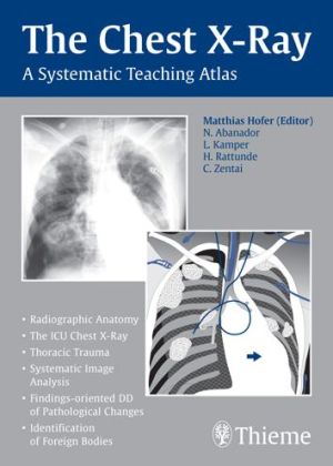
The Chest X-Ray: A Systematic Teaching Atlas
Pages: 224,
Specialty: Radiology,
Publisher: Thieme,
Publication Year: 2006,
Cover: Paperback,
Dimensions: 214x300x12mm
For whom is this book designed? For all students and physicians in training who want to learn more about the systematic interpretation of conventional chest radiographs, and for anyone who wants to learn how to insert chest tubes and central venous catheters. What does this book offer? * Detailed diagrams on topographical anatomy, with numerical labels for self-review.* Coverage includes even relatively complex findings in trauma victims and ICU patients.* Detailed, step-by-step instructions on the placement of CVCs and chest tubes.* Simple aids and tricks, such as the "silhouette sign," that are helpful in image interpretation.* Images to illustrate all common abnormalities (systematically arranged according to morphological patterns).show more
Specialty: Radiology,
Publisher: Thieme,
Publication Year: 2006,
Cover: Paperback,
Dimensions: 214x300x12mm
For whom is this book designed? For all students and physicians in training who want to learn more about the systematic interpretation of conventional chest radiographs, and for anyone who wants to learn how to insert chest tubes and central venous catheters. What does this book offer? * Detailed diagrams on topographical anatomy, with numerical labels for self-review.* Coverage includes even relatively complex findings in trauma victims and ICU patients.* Detailed, step-by-step instructions on the placement of CVCs and chest tubes.* Simple aids and tricks, such as the "silhouette sign," that are helpful in image interpretation.* Images to illustrate all common abnormalities (systematically arranged according to morphological patterns).show more
Product Information
Product Information
Shipping & Returns
Shipping & Returns



The Chest X-Ray: A Systematic Teaching Atlas
The Chest X-Ray: A Systematic Teaching Atlas
Pages: 224,
Specialty: Radiology,
Publisher: Thieme,
Publication Year: 2006,
Cover: Paperback,
Dimensions: 214x300x12mm
For whom is this book designed? For all students and physicians in training who want to learn more about the systematic interpretation of conventional chest radiographs, and for anyone who wants to learn how to insert chest tubes and central venous catheters. What does this book offer? * Detailed diagrams on topographical anatomy, with numerical labels for self-review.* Coverage includes even relatively complex findings in trauma victims and ICU patients.* Detailed, step-by-step instructions on the placement of CVCs and chest tubes.* Simple aids and tricks, such as the "silhouette sign," that are helpful in image interpretation.* Images to illustrate all common abnormalities (systematically arranged according to morphological patterns).show more
Specialty: Radiology,
Publisher: Thieme,
Publication Year: 2006,
Cover: Paperback,
Dimensions: 214x300x12mm
For whom is this book designed? For all students and physicians in training who want to learn more about the systematic interpretation of conventional chest radiographs, and for anyone who wants to learn how to insert chest tubes and central venous catheters. What does this book offer? * Detailed diagrams on topographical anatomy, with numerical labels for self-review.* Coverage includes even relatively complex findings in trauma victims and ICU patients.* Detailed, step-by-step instructions on the placement of CVCs and chest tubes.* Simple aids and tricks, such as the "silhouette sign," that are helpful in image interpretation.* Images to illustrate all common abnormalities (systematically arranged according to morphological patterns).show more
$31.50
The Chest X-Ray: A Systematic Teaching Atlas—
$31.50
Product Information
Product Information
Shipping & Returns
Shipping & Returns
Description
Pages: 224,
Specialty: Radiology,
Publisher: Thieme,
Publication Year: 2006,
Cover: Paperback,
Dimensions: 214x300x12mm
For whom is this book designed? For all students and physicians in training who want to learn more about the systematic interpretation of conventional chest radiographs, and for anyone who wants to learn how to insert chest tubes and central venous catheters. What does this book offer? * Detailed diagrams on topographical anatomy, with numerical labels for self-review.* Coverage includes even relatively complex findings in trauma victims and ICU patients.* Detailed, step-by-step instructions on the placement of CVCs and chest tubes.* Simple aids and tricks, such as the "silhouette sign," that are helpful in image interpretation.* Images to illustrate all common abnormalities (systematically arranged according to morphological patterns).show more
Specialty: Radiology,
Publisher: Thieme,
Publication Year: 2006,
Cover: Paperback,
Dimensions: 214x300x12mm
For whom is this book designed? For all students and physicians in training who want to learn more about the systematic interpretation of conventional chest radiographs, and for anyone who wants to learn how to insert chest tubes and central venous catheters. What does this book offer? * Detailed diagrams on topographical anatomy, with numerical labels for self-review.* Coverage includes even relatively complex findings in trauma victims and ICU patients.* Detailed, step-by-step instructions on the placement of CVCs and chest tubes.* Simple aids and tricks, such as the "silhouette sign," that are helpful in image interpretation.* Images to illustrate all common abnormalities (systematically arranged according to morphological patterns).show more